

An electroretinogram (ERG) measures the electrical signals produced by the retina in response to light flashes. It evaluates the function of photoreceptors (rods and cones) and other retinal cells.
Key takeaways
- Measures retinal function in response to light
- Evaluates rods and cones (photoreceptors)
- Helpful for unexplained vision loss
- Diagnoses retinal dystrophies
- May detect cancer-associated retinopathy
Questions about your diagnosis? Contact our office
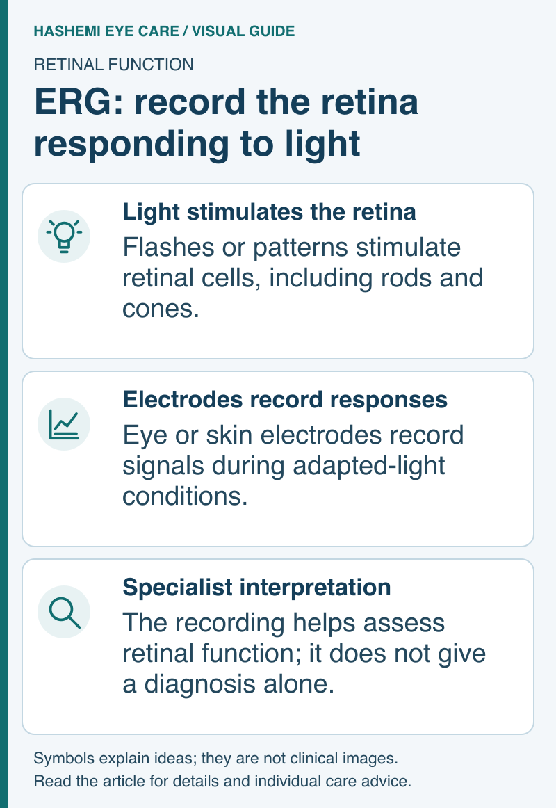
Visual guide: ERG: record the retina responding to light
Read this visual as text
Light stimulates the retina: Flashes or patterns stimulate retinal cells, including rods and cones. Electrodes record responses: Eye or skin electrodes record signals during adapted-light conditions. Specialist interpretation: The recording helps assess retinal function; it does not give a diagnosis alone.
Why It's Done
- Diagnose inherited retinal dystrophies (retinitis pigmentosa)
- Evaluate unexplained vision or night vision loss
- Detect cancer-associated retinopathy
- Assess retinal toxicity from medications
- Differentiate retinal from optic nerve disease
What to Expect
Full-Field ERG
Before:
- Sit in dark room for 20-30 minutes (dark adaptation)
- Dilating drops may be used
During:
- Contact lens electrode or skin electrodes placed
- Light flashes presented
- Response recorded
- Both dark-adapted and light-adapted testing
- Takes 45-60 minutes
Multifocal ERG (mfERG)
- Tests small areas of central retina
- Hexagonal pattern of stimuli
- Takes 30-45 minutes
- More detailed central retinal map
Getting Home After Testing
If dilating drops are used, bring sunglasses and arrange a ride if your vision is blurred or light-sensitive. Wait until vision is clear and comfortable before driving. Ask the testing team when to expect the specialist's interpretation; the recording alone does not give a diagnosis.
What It Shows
Normal ERG
- Good responses from rods (dim light) and cones (bright light)
- Normal waveform shape and timing
Abnormal Findings
- Reduced responses-retinal dysfunction
- Absent rod responses-rod-cone dystrophy
- Negative ERG-specific pattern suggests certain conditions
ERG vs VEP
| ERG | VEP |
|---|---|
| Tests retina | Tests visual pathway to brain |
| Flash stimulus | Pattern stimulus |
| Eye electrodes | Scalp electrodes |
| Retinal disease | Optic nerve/brain disease |
References
- American Academy of Ophthalmology. Electroretinogram.
- National Eye Institute. Retinitis Pigmentosa.
Medical Disclaimer: This information is for educational purposes only and does not replace professional medical advice.