

Optic neuritis often presents with vision loss in one eye over hours to days, reduced color intensity, and pain with eye movement. The inflamed optic nerve carries visual information from the eye to the brain. Many typical cases improve, but the cause is important. MS-related optic neuritis, neuromyelitis optica (NMO), MOG antibody disease, infection, and other inflammatory diseases can differ substantially in prognosis and treatment.
Key takeaways
- Vision loss usually builds over hours to days, most often in one eye
- Eye-movement pain is a major diagnostic feature, especially when color vision is reduced
- Typical optic neuritis often improves, but recovery speed and treatment depend on the cause
- MS, NMO, and MOGAD have different clinical implications and are not interchangeable labels
- MRI is central for diagnosis, MS-risk assessment, and treatment decisions
- New vision loss needs same-day triage; bilateral, severe, or rapidly worsening loss needs emergency assessment
Questions about your diagnosis? Contact our office
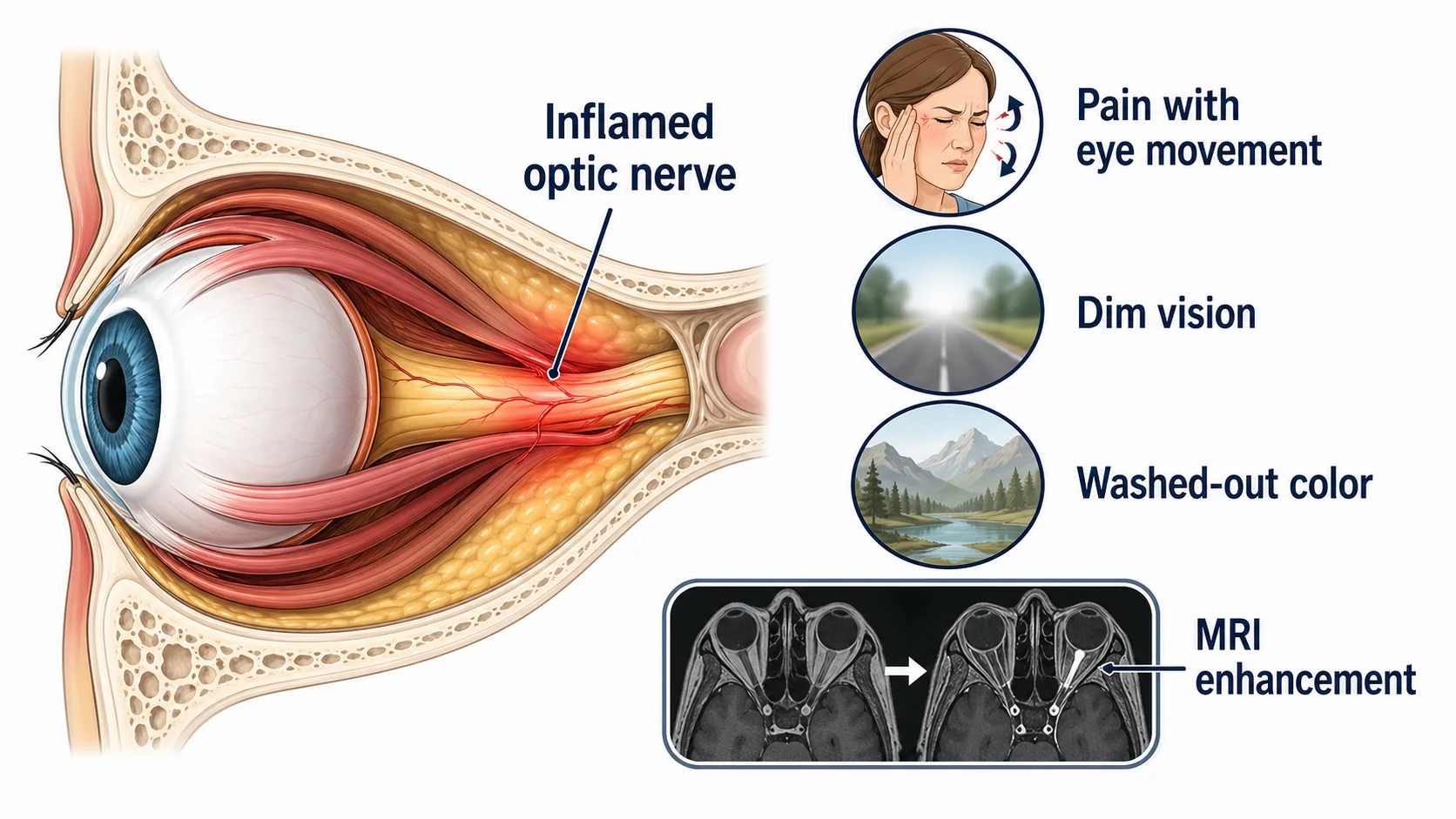
What Optic Neuritis Feels Like
Typical symptoms include:
- Vision loss developing over hours to days
- Usually affects one eye at a time
- Vision may be blurry, dim, or have a "film" over it
- Pain with eye movement: characteristic symptom
- Pain may precede vision loss by a few days
- Color vision changes: red appears washed out
- Vision worse with heat or exercise (Uhthoff phenomenon)
- Flashing lights with eye movement (phosphenes)
What Causes Optic Neuritis
Demyelinating Disease (Most Common)
Multiple Sclerosis (MS)
- Optic neuritis is first symptom in ~20% of MS patients
- In the ONTT cohort of typical acute optic neuritis, about 50% developed MS within 15 years; this estimate does not apply to every cause of optic nerve inflammation
- MRI findings help predict MS risk
Neuromyelitis Optica (NMO/Devic's disease)
- Autoimmune attack on optic nerve and spinal cord
- Often more severe than MS-related optic neuritis
- AQP4 antibody positive
- Higher risk of poor recovery
MOG Antibody Disease (MOGAD)
- Autoimmune condition with MOG antibodies
- Can cause severe but often recoverable optic neuritis
- May be bilateral
- Different treatment approach than MS
Other Causes
- Infections (viral, Lyme disease, syphilis, cat-scratch disease)
- Inflammatory conditions (sarcoidosis, lupus)
- Vaccination-related (rare)
- Idiopathic (no cause found)
When to Seek Care
Go to the emergency department the same day if vision loss is bilateral, severe, rapidly worsening, or accompanied by new neurologic symptoms. Call 911 immediately for sudden vision loss, even without pain: central retinal artery occlusion (CRAO) can resemble other optic-nerve problems. Also call 911 for BE-FAST stroke symptoms, confusion, trouble walking, or sudden severe headache.
Contact ophthalmology or neuro-ophthalmology the same day for triage of:
- Eye pain, especially with eye movement
- One-eye vision loss that worsens over hours to days
- Washed-out color vision in one eye
How Optic Neuritis Is Diagnosed
Clinical Examination
- Visual acuity: usually reduced
- Color vision: red desaturation characteristic
- Pupil exam: relative afferent pupillary defect (RAPD) present
- Visual field: various patterns of loss
- Fundus exam: optic disc may look normal or swollen
Key Tests
-
MRI brain and orbits with contrast
- Shows optic nerve inflammation
- Brain lesions predict MS risk
- Essential for diagnosis and prognosis
-
Optical coherence tomography (OCT)
- Measures nerve fiber layer thickness
- May show swelling acutely
- Thinning develops over months
-
Visual evoked potentials (VEP)
- Shows delayed signal conduction
- Can remain abnormal even after visual recovery
-
- NMO-IgG (AQP4 antibodies)
- MOG antibodies
- Other tests based on suspected causes
-
Lumbar puncture: sometimes needed
Treatment
Observation
For typical optic neuritis:
- Vision often improves on its own over 2-12 weeks
- In the Optic Neuritis Treatment Trial, even among eyes starting at 20/200 or worse, about 85% improved to 20/40 or better by 6 months
- Treatment may not change final outcome but speeds recovery
IV Methylprednisolone
When used:
- Severe vision loss
- Pain that is interfering with sleep, work, or basic daily tasks
- Need faster recovery (work, bilateral involvement)
Protocol:
- Typically 1 gram IV daily for 3-5 days
- May be followed by oral steroids (taper)
- Speeds recovery but may not change final outcome
The dose matters: The ONTT found increased recurrence with low-dose oral prednisone alone (1 mg/kg/day). That finding does not apply to every oral regimen. A later randomized trial found no significant recovery difference between bioequivalent high-dose oral and IV treatment in acute demyelinating optic neuritis. Your specialist chooses the route and dose; do not use leftover steroids or substitute one regimen for another.
For NMO/MOGAD
More aggressive treatment needed:
- High-dose IV steroids
- Plasmapheresis if severe or not responding
- Relapse-prevention treatment is central in AQP4-positive NMO; in MOGAD, the long-term plan depends on attack severity, recurrence, and specialist assessment
Prognosis and Recovery
Visual Recovery
- Typical demyelinating optic neuritis often has substantial visual recovery; ONTT data found about 85% of eyes starting at 20/200 or worse improved to 20/40 or better by 6 months, but atypical causes can have different treatment needs and prognosis
- Recovery usually begins within 2-4 weeks
- Continues for several months
- Some color or contrast vision deficit may persist
- Vision may temporarily worsen with heat/exercise (Uhthoff phenomenon)
Risk of Multiple Sclerosis
For detailed information about the connection between optic neuritis and MS, including what MRI findings mean for your risk and treatment decisions, see our guide: Optic Neuritis and MS Risk - Understanding Your Diagnosis.
The following 15-year estimates come from the ONTT cohort of typical acute optic neuritis, rather than all optic neuritis diagnoses. MRI brain findings helped predict risk:
| MRI Finding | 15-Year MS Risk |
|---|---|
| Normal MRI (no lesions) | ~25% |
| 1 or more brain lesions | ~72% |
Other factors affecting risk:
- Female sex
- Younger age
- Presence of oligoclonal bands in spinal fluid
Recurrence
- About 35% have another episode of optic neuritis within 10 years
- May affect same or opposite eye
- Consider disease-modifying treatment if MS or NMO diagnosed
Living with Optic Neuritis
During Acute Phase
- Rest your eyes when fatigued
- Use good lighting
- Avoid excessive heat (hot showers, saunas)
- Pain medication as needed
- Plan for reduced activities temporarily
After Recovery
- Regular neuro-ophthalmology and/or neurology follow-up
- MRI surveillance if MS risk factors present
- Monitor for new symptoms
- Consider disease-modifying therapy if indicated
Frequently Asked Questions
Will I go blind from optic neuritis?
Typical MS-associated optic neuritis often recovers useful vision, though color and contrast deficits may persist. NMO-associated and other atypical forms can cause severe permanent loss, so prognosis depends on the cause and prompt treatment.
Does optic neuritis mean I have MS?
Not necessarily. About half of the ONTT participants with typical acute optic neuritis developed MS over 15 years. That estimate does not apply to MOGAD, NMO, or every cause of optic neuritis. MRI and other findings guide your individual assessment.
Why does my vision get worse when I'm hot?
This is called Uhthoff phenomenon. Demyelinated nerves conduct signals more slowly when warm. It's temporary-vision returns to baseline when you cool down. It doesn't mean optic neuritis is recurring.
Can optic neuritis come back?
Yes, recurrence is possible in about 35% of cases within 10 years. It may affect the same eye or the other eye. Recurrent optic neuritis increases suspicion for MS, NMO, or MOGAD.
What makes optic neuritis "atypical"?
Bilateral onset, very severe vision loss, poor early recovery, marked optic disc swelling with hemorrhages, recurrent attacks, older age, lack of eye-movement pain, or prominent neurologic/systemic symptoms can suggest NMO, MOGAD, infection, sarcoidosis, ischemia, or another mimic. Those patterns usually need broader blood testing and more urgent treatment decisions than typical MS-associated optic neuritis.
Should I start MS treatment?
This decision depends on MRI findings, presence of oligoclonal bands, and individual risk factors. If MS risk is high, early treatment may reduce future attacks. Discuss with your neurologist.
Can I drive with optic neuritis?
During acute vision loss, likely not. After recovery, it depends on your visual acuity and field. Discuss driving restrictions with your doctor.
References
-
American Heart Association. CRAO emergency management.
-
Optic Neuritis Study Group. Multiple sclerosis risk after optic neuritis: final optic neuritis treatment trial follow-up. Arch Neurol. 2008;65(6):727-732.
-
Beck RW, Cleary PA, Backlund JC. The course of visual recovery after optic neuritis: experience of the Optic Neuritis Treatment Trial. Ophthalmology. 1994;101(11):1771-1778.
-
Morrow SA, et al. Effect of treating acute optic neuritis with bioequivalent oral vs intravenous corticosteroids: a randomized clinical trial. JAMA Neurol. 2018;75(6):690-696.
-
Petzold A, et al. Diagnosis and classification of optic neuritis. Lancet Neurol. 2022;21(12):1120-1134.
-
North American Neuro-Ophthalmology Society. Optic Neuritis.
-
StatPearls. Optic Neuritis. NCBI Bookshelf.
Medical Disclaimer: This information is for educational purposes only and does not replace professional medical advice, diagnosis, or treatment. If you have concerns about optic neuritis or any symptoms, please consult a qualified healthcare provider.