Testing library
Diagnostic Tests
Understand why a test is ordered, how to prepare, and what the results can tell you.
39 test guides
Browse 5 categories
Related: Conditions these tests diagnose • Clinical findings
39 articles. Filter by a word or phrase.
Amsler Grid Testing
A simple home monitoring tool for detecting macular problems. Learn how to use the Amsler grid to catch vision changes early.
Biometry (IOL Calculation)
Precise eye measurements for calculating intraocular lens power before cataract surgery. Learn how this test helps choose a lens-power target.
Color Vision Testing
Tests to evaluate your ability to distinguish colors, which can be affected by optic nerve and retinal conditions.
Contrast Sensitivity Testing
A test measuring your ability to distinguish objects from their background, often affected in optic nerve conditions.
Exophthalmometry (Hertel)
A bedside measurement of how far the eyes protrude from the orbit. Critical for monitoring thyroid eye disease and orbital tumors.
Eye Movement Examination
Assessment of how the eyes move together, essential for diagnosing causes of double vision and eye misalignment.
Fundoscopic Examination
Direct visualization of the back of the eye (retina, optic nerve, blood vessels) using an ophthalmoscope or other instruments.
Gonioscopy
An examination of the eye's drainage angle using a special lens. Essential for diagnosing and classifying glaucoma.
Hess Chart Test
A specialized test that maps how each eye moves into the nine cardinal positions, used to characterize cranial nerve palsies and other motility disorders.
Ice Test for Myasthenia Gravis
A simple bedside test using ice on the eyelid to help diagnose myasthenia gravis.
Maddox Rod Test
A simple bedside test that uses a striped lens to dissociate the eyes and quantify the direction and amount of double vision.
Pachymetry (Corneal Thickness Measurement)
A test measuring corneal thickness, important for glaucoma assessment and refractive surgery planning. Learn what to expect and what results mean.
Prism and Cover Test
A clinical test using prisms to measure eye misalignment and determine the cause of double vision.
Pupil Examination
Assessment of pupil size, shape, and reactions that can reveal important information about the visual and nervous system.
Slit Lamp Examination
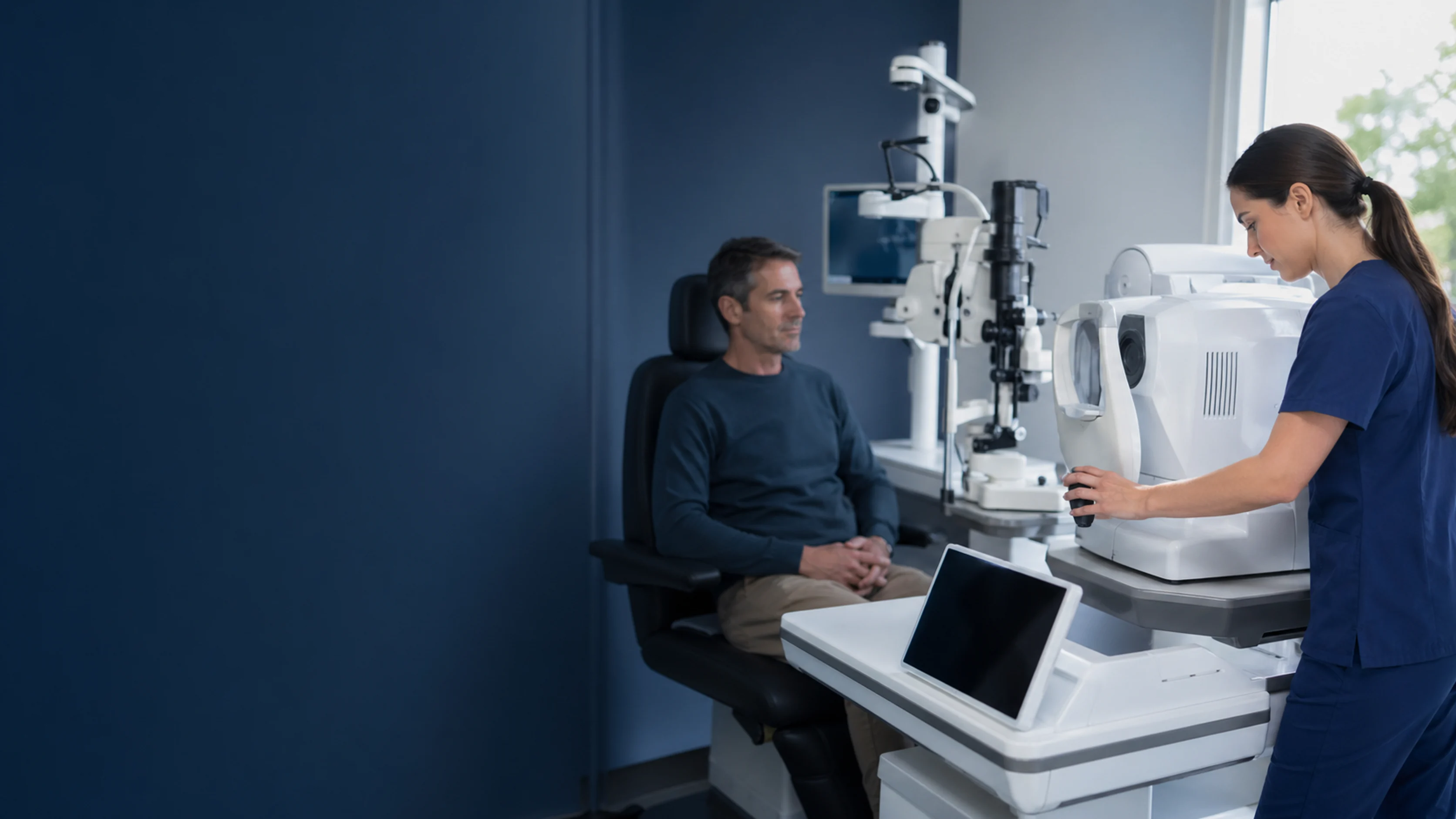
A detailed examination of the eye structures using a specialized microscope with adjustable light, allowing magnified views of the eye's front and back.
Tonometry (Eye Pressure Measurement)
A test to measure the pressure inside your eye. Learn about different types of tonometry and why it's essential for glaucoma screening.
Visual Field Test (Perimetry)
A test that maps your peripheral and central vision to detect blind spots and patterns of vision loss.
Worth Four-Dot Test
A simple binocular vision test using red-green glasses and four colored lights to assess fusion, suppression, and diplopia.
Anterior Segment OCT
A non-contact light-based imaging method that produces detailed cross-sections of the cornea, anterior chamber, and angle. Often used in glaucoma and corneal evaluation.
B-Scan Ultrasound
An imaging test that uses sound waves to visualize the structures inside the eye when the view is blocked by blood, cataracts, or other opacities.
Carotid Ultrasound (Doppler)
An ultrasound test to evaluate blood flow in the neck arteries that supply the eyes and brain.
Corneal Topography
A non-invasive imaging test that maps the curvature and shape of the cornea to diagnose astigmatism, keratoconus, and other corneal conditions.
CT Scan (Head/Orbits)
A quick imaging test using X-rays to visualize the brain, orbits, and bony structures.
Fluorescein Angiography
An imaging test using injected dye to evaluate blood flow in the retina and detect leakage or abnormal vessels.
Fundus Autofluorescence (FAF)
A non-invasive imaging test that detects metabolic changes in the retina by capturing the natural fluorescence of the retinal pigment epithelium.
Fundus Photography
Photographs of the back of the eye (retina and optic nerve) to document and monitor eye conditions.
MRI Brain and Orbits
Magnetic resonance imaging of the brain and eye sockets to evaluate the visual pathways, optic nerves, and surrounding structures.
OCT Angiography (OCTA)
Advanced OCT imaging that visualizes blood vessels in the retina and optic nerve without injecting dye.
OCT (Optical Coherence Tomography)
A non-invasive imaging scan that provides detailed cross-sectional images of the retina and optic nerve.
Ultrasound Biomicroscopy (UBM)
A high-frequency ultrasound that produces detailed images of the front of the eye, especially the angle, ciliary body, and structures behind the iris.
ERG (Electroretinogram)
A test that measures the electrical responses of the retina to light, helping diagnose retinal diseases.
Single-Fiber EMG
A specialized nerve and muscle test that is highly sensitive for detecting myasthenia gravis, particularly when other tests are negative.
Visual Evoked Potential (VEP)
A test that measures the electrical response of your brain to visual stimulation, detecting problems in the visual pathway.
Acetylcholine Receptor Antibody Test
A blood test that detects antibodies associated with myasthenia gravis, helping support the diagnosis of this neuromuscular condition.
Autoantibody Testing Panels
Blood tests that detect antibodies against the body's own tissues, used to diagnose autoimmune conditions affecting the eyes and nervous system.
Blood Tests for Neuro-Ophthalmic Conditions
Common blood tests ordered to help diagnose the underlying cause of visual and neurological symptoms.
Genetic Testing for Eye Conditions
DNA testing to diagnose inherited conditions affecting vision, including LHON and dominant optic atrophy.