

Fluorescein angiography (FA) is an imaging test where fluorescent dye is injected into a vein, and photographs are taken as the dye travels through the blood vessels in the eye. It reveals blood flow patterns, leakage, and vascular abnormalities.
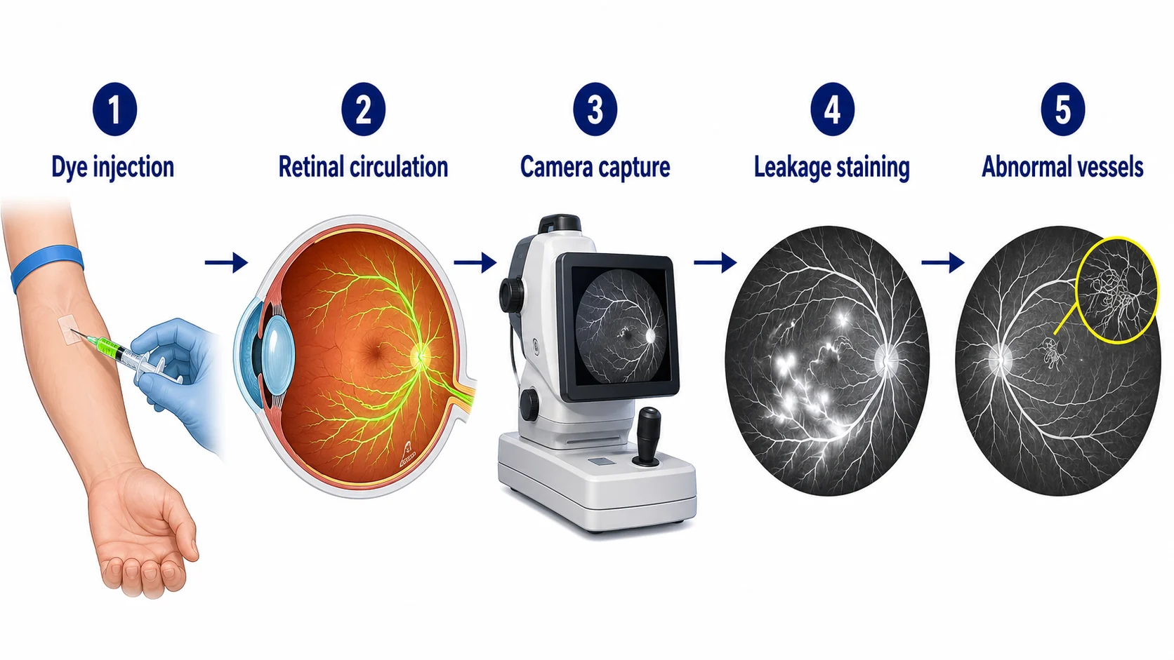
Key takeaways
- Dye injected into arm vein
- Photographs blood flow in eye
- Detects leakage and abnormal vessels
- Skin and urine turn yellow temporarily
- Important for diagnosing vascular conditions
Questions about your diagnosis? Contact our office
Why It's Done
- Evaluate optic disc swelling vs pseudopapilledema
- Diagnose ischemic optic neuropathy
- Assess retinal vascular diseases
- Look for inflammation (vasculitis)
- Guide treatment decisions
What to Expect
Before the Test
- Pupils dilated
- IV line placed in an arm vein
- Tell the staff about any prior reaction to fluorescein or contrast dye, severe allergies, asthma, kidney disease, or pregnancy
- Pregnancy is generally a relative contraindication; the test is usually deferred unless absolutely necessary
- If indocyanine green (ICG) angiography is planned too, it uses a different dye with separate precautions. Tell the team about prior dye reactions, severe allergies, and liver disease. Shellfish allergy alone does not predict a dye reaction.
During the Test
- Fluorescein dye injected through the IV
- Rapid sequence of photos taken
- Bright flashes of light
- Photos continue for several minutes
- Takes 15-30 minutes total
After the Test
- Skin and urine appear yellow-orange for 24-48 hours
- This is expected and temporary
- Follow your usual fluid plan and any instructions from the clinic; keep existing fluid restrictions
- Bring sunglasses after dilation. Arrange a ride if vision is blurred or light-sensitive, and wait for clear comfortable vision before driving
Risks and Side Effects
Common (mild)
- Yellow discoloration of skin and urine
- Brief nausea or warm flushing during injection
- Mild itching
Less common
- Vomiting
- Hives (urticaria)
- Extravasation - if dye leaks out of the vein, it can cause local pain, skin staining, and rarely tissue necrosis. Tell the technician immediately if you feel a sudden burning at the IV site.
Rare but serious
- Anaphylaxis and severe allergic reactions including bronchospasm, throat swelling, hypotension, and cardiac arrest. Severe reactions occur in roughly 1 in 1,900 patients; deaths from fluorescein anaphylaxis are very rare (estimated about 1 in 220,000). Photography rooms should have resuscitation equipment available and staff trained to use it.
- Vasovagal syncope (fainting)
- Myocardial infarction or cardiac arrhythmia in patients with significant cardiac disease
If you have ever had a serious reaction to fluorescein or other IV contrast, tell your team - alternative imaging (such as OCT angiography, which does not use injected dye) can often answer the same clinical question.
What It Shows
Normal
- Even filling of vessels
- No leakage
Abnormal
- Disc leakage-inflammation or papilledema
- Delayed filling-poor blood flow
- Hyperfluorescence-drusen, scars
- Blocked fluorescence-hemorrhage
Neuro-Ophthalmic Uses
- Distinguish true disc edema from drusen
- Evaluate optic nerve ischemia
- Assess inflammatory conditions
References
- American Academy of Ophthalmology. Fluorescein Angiography.
- National Eye Institute. Age-Related Macular Degeneration.
Medical Disclaimer: This information is for educational purposes only and does not replace professional medical advice.