

Scleral buckle surgery is a long-established technique for repairing retinal detachment. The buckle element is either a solid silicone band (typically used as an encircling 360° element around the eye) or a softer silicone sponge (typically placed segmentally over the area of the retinal tear); many surgeries use both an encircling band and a segmental sponge. The buckle is sutured to the outside of the eye, creating an indentation that pushes the wall of the eye toward the detached retina. The indentation supports retinal tears and reduces vitreous traction, allowing the retina to reattach. Buckle surgery is often combined with vitrectomy for complex cases and is sometimes performed alone for selected primary detachments.
Key takeaways
- Scleral buckle places silicone support on the outside of the eye to support the retina and close tears
- Single-procedure success rates are approximately 85-92% for primary rhegmatogenous retinal detachment in appropriate cases
- Particularly suited to younger phakic patients with simple detachments and tears in the lower retina
- Combined buckle plus vitrectomy is common in complex detachments
- Recovery includes mild discomfort, eye redness, and a typical myopic shift of approximately 1-3 diopters (sometimes more) when an encircling element is used; segmental sponges cause less shift
Questions about your diagnosis? Contact our office
Visual guide: Scleral buckle for retinal detachment
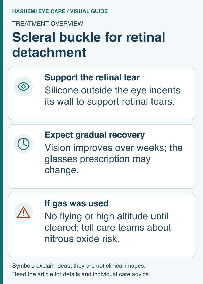
Read this visual as text
When Scleral Buckle Is Used
- Primary rhegmatogenous retinal detachment - particularly with inferior tears that pneumatic retinopexy cannot reach
- Young phakic patients - buckle does not accelerate cataract as much as vitrectomy
- Recurrent detachment after pneumatic retinopexy or initial vitrectomy
- Combined with vitrectomy in complex or proliferative vitreoretinopathy cases
How the Procedure Works
- Performed in the operating room under local or general anesthesia
- Conjunctiva is opened and extraocular muscles are isolated
- A flexible silicone band or sponge segment is sutured to the sclera
- The buckle creates a targeted or circumferential indentation of the eye wall
- Retinal tears are sealed with cryotherapy or laser
- Subretinal fluid is sometimes drained
- Conjunctiva is closed; the band remains permanently in place
Recovery
- Hospital stay typically not required
- Eye may be patched immediately after surgery
- Mild to moderate discomfort for several days, controlled with oral analgesics
- Eye redness and bruising on the conjunctival surface
- Vision improves over weeks as the retina settles
- A myopic shift in glasses prescription of about 1-3 D commonly occurs with an encircling buckle because the buckle elongates the eye's axial length; segmental (non-encircling) sponges produce less shift
- Postoperative drops often include a topical antibiotic briefly and a steroid taper
- Most patients can return to work in 1-2 weeks
Ask whether a gas bubble was used with your buckle or combined vitrectomy. If so, follow the gas-specific positioning and travel restrictions:
If air or gas is inside your eye, do not fly or travel to high altitude until your surgeon confirms it is safe. Tell every anesthesia, dental, emergency, and ambulance team before they give you treatment: nitrous oxide, including Entonox or other "gas and air," can expand the bubble and cause severe vision loss. Ask for a gas-warning card or wristband and keep it with you until the bubble is gone.
Call the surgeon immediately for increasing pain, worsening vision, discharge, or new flashes, floaters, or a curtain in your sight. Seek emergency eye care if the team cannot be reached.
Risks
- Infection - uncommon
- Diplopia - from disturbance of extraocular muscles; usually transient
- Refractive shift - myopic shift, typically 1-3 diopters with encircling elements (segmental sponges produce less)
- Buckle exposure or extrusion - rare; may require removal years later
- Anterior segment ischemia - rare; most often associated with extensive disinsertion of multiple rectus muscles during buckle placement, which compromises the anterior ciliary artery blood supply. More common in patients with sickle cell disease or other vascular comorbidities
- Recurrent detachment - 10-20% require additional surgery
- Cataract progression - slower than after vitrectomy
Scleral Buckle vs. Vitrectomy vs. Pneumatic Retinopexy
| Feature | Scleral buckle | Vitrectomy | Pneumatic retinopexy |
|---|---|---|---|
| Setting | OR | OR | Office |
| Anatomical success | 85-92% | 85-95% | ~75-80% |
| Cataract progression | Slow | Faster | Slowest |
| Post-op refractive change | Mild myopic shift | Minimal | Minimal |
| Ideal for | Young phakic patients, lower-retina tears | Complex cases, pseudophakic patients | Single upper-retina tear, cooperative patient |
Frequently Asked Questions
Will the silicone band stay in my eye forever?
Usually yes. The band is buried under the conjunctiva and is generally well tolerated for life. In rare cases, such as exposure, infection, or persistent diplopia, the band can be removed years later.
Why might I need a buckle and a vitrectomy together?
Some complex detachments - particularly those with proliferative vitreoretinopathy, giant tears, or extensive retinal involvement - benefit from both techniques. The buckle provides ongoing scleral support; the vitrectomy clears tractional vitreous and allows direct retinal reattachment.
Will my vision come back to normal?
That depends on how long the macula was detached and how complete the reattachment is. Macula-on detachments treated promptly usually recover good vision; macula-off detachments often have some permanent reduction in central acuity even after successful reattachment.
References
-
Royal College of Ophthalmologists. Safety Alert: Do Not Use Nitrous Oxide When There Is Gas in an Operated Eye.
-
Heimann H, Bartz-Schmidt KU, Bornfeld N, et al; Scleral Buckling versus Primary Vitrectomy in Rhegmatogenous Retinal Detachment Study Group. Scleral buckling versus primary vitrectomy in rhegmatogenous retinal detachment: a prospective randomized multicenter clinical study. Ophthalmology. 2007;114(12):2142-2154.
-
American Academy of Ophthalmology EyeWiki. Scleral Buckle.
Medical Disclaimer: This information is for educational purposes only and does not replace professional medical advice.