
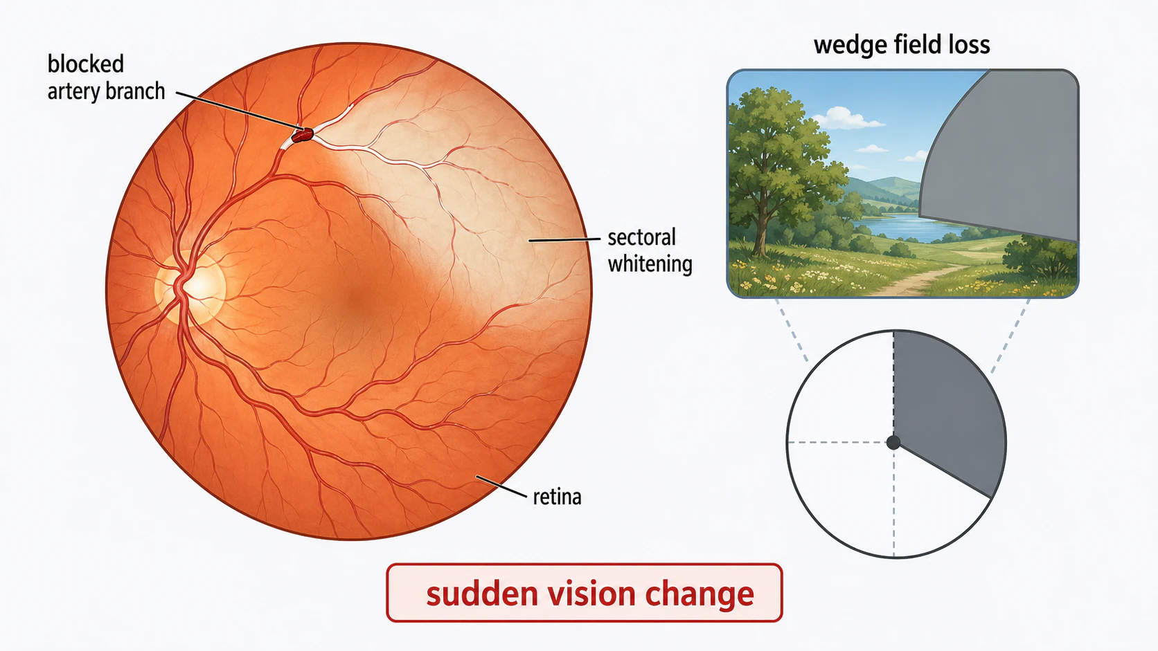
Branch retinal artery occlusion (BRAO) occurs when one of the smaller arteries supplying blood to a portion of the retina becomes blocked, usually by an embolus (a piece of material that traveled from elsewhere). It causes sudden, painless vision loss affecting part of the visual field. Like central retinal artery occlusion (CRAO), BRAO is a warning sign of stroke risk.
Key takeaways
- Branch artery blocked: affects a portion of the retina
- Sudden, painless vision loss in part of the visual field
- Usually caused by embolus from carotid artery or heart
- Better visual prognosis than CRAO: often some vision preserved
- Stroke warning sign: urgent cardiovascular and carotid workup needed
- Damaged retina usually does not recover; focus is on preventing stroke and protecting remaining vision
Questions about your diagnosis? Contact our office
Call 911 now for a sudden missing area or loss of vision in one eye, even if it clears. Do not drive yourself or wait for a routine eye appointment. Acute retinal artery occlusion needs emergency stroke assessment; note when symptoms began.
What Happens in BRAO
The Branch Retinal Arteries
- Branch off from the central retinal artery
- Supply blood to different sectors of the retina
- When blocked, that sector loses blood supply
Consequences
- Immediate: affected retinal area becomes ischemic (oxygen-starved)
- The retina in that area is damaged or dies
- Creates a blind spot (scotoma) in the corresponding visual field
- Unaffected areas retain normal function
Causes
Emboli (Most Common)
Material that breaks off from elsewhere and lodges in the artery:
Cholesterol Emboli (Hollenhorst Plaques):
- From atherosclerotic plaque in carotid artery
- Appear as shiny, yellow-orange crystals in the artery
- Most common type
Fibrin-Platelet Emboli:
- Grayish-white, elongated
- From carotid or cardiac source
- May move or break up
Calcific Emboli:
- White, from calcified heart valves
- Don't move (embedded in vessel)
- Less common
Sources of Emboli
Carotid Artery:
- Atherosclerotic plaque
- Most common source
- Same risk as for stroke
Heart:
- Atrial fibrillation
- Valve disease
- Recent heart attack
- Endocarditis
Other:
- Aortic arch
- Fat emboli (after trauma or surgery)
- Talc (in IV drug users)
Other Causes
- Giant cell arteritis (consider urgently in patients over 50, especially with headache, jaw pain, scalp tenderness, fever, or systemic symptoms)
- Vasculitis
- Hypercoagulable states
- Vasospasm
Symptoms
Classic Presentation
- Sudden vision loss: affects part of the visual field
- Painless
- Described as "a spot is missing" or "part of my vision is gone"
- Often noted immediately (unlike gradual losses)
Visual Field Defect
The visual field loss corresponds to the retinal area affected:
- Usually an arcuate (curved) or wedge-shaped defect
- Altitudinal defect (upper or lower field loss) common
- Central vision may be preserved if fovea is spared
Transient BRAO
Some patients experience transient episodes:
- Vision loss lasting minutes, then resolving
- Called "amaurosis fugax" (affecting part of vision)
- Warning sign of impending permanent occlusion
- Requires urgent evaluation
Diagnosis
Clinical Examination
Fundus Findings:
- Pale, whitened retina in the affected sector
- Visible embolus at a branch point (sometimes)
- "Box-carring" of blood in affected artery (segmented column)
- May see cherry-red spot if macular branch affected
Visual Field Testing
- Documents the scotoma (blind spot)
- Helps correlate with fundus findings
- Useful for monitoring
Imaging
- Shows blocked vessel and delayed filling
- Delineates extent of non-perfusion
OCT:
- Shows retinal thickening acutely (edema)
- Retinal thinning later (atrophy)
Critical Workup
BRAO indicates high stroke risk. Urgent workup includes:
- Carotid ultrasound or CTA (looking for plaque)
- Echocardiogram (looking for cardiac source)
- ECG (looking for atrial fibrillation)
- Blood pressure
- Blood glucose, lipid panel
- ESR and CRP to assess for giant cell arteritis, especially after age 50; normal results do not exclude it or justify delaying treatment when clinical suspicion is high
- Consider MRI/MRA brain
Treatment
For the Eye
Emergency assessment comes first. Do not massage the eye or try pressure-lowering treatment at home. The stroke and ophthalmology teams assess whether any acute intervention is appropriate; options to restore established retinal damage are limited. Identifying the source and preventing another retinal event or brain stroke remain central.
Systemic Treatment (Critical)
Stroke Prevention:
- Aspirin or other antiplatelet therapy
- Statin therapy for cholesterol
- Blood pressure control
- Diabetes management
If Carotid Disease Found:
- Carotid endarterectomy (surgery) for significant stenosis
- May be more urgent than for asymptomatic disease
If Cardiac Source Found:
- Anticoagulation for atrial fibrillation
- Valve evaluation if indicated
If Giant Cell Arteritis Suspected:
- Immediate high-dose steroids
- Can prevent bilateral blindness and stroke
Prognosis
Visual Outcome
Generally Better Than CRAO:
- Unaffected retina maintains function
- Central vision often preserved (if fovea spared)
- Many patients retain useful vision
- Some natural improvement as edema resolves
Permanent Defect:
- The scotoma (blind spot) usually remains
- Some patients adapt well
- Central vision preservation is key to function
Systemic Prognosis
BRAO patients have increased risk of:
- Stroke
- Heart attack
- Cardiovascular death
This is why cardiovascular workup and prevention are so important. The retinal event is a warning sign.
Comparison: BRAO vs. CRAO
| Feature | BRAO | CRAO |
|---|---|---|
| Area affected | Sector of retina | Entire retina |
| Vision loss | Partial field | Severe, whole eye |
| Prognosis | Often good functional vision | Usually poor |
| Stroke risk | Elevated | Elevated |
| Emergency level | Emergency stroke assessment | Emergent |
Follow-Up
Acute Phase
- Urgent cardiovascular workup
- Repeat exam in 1-2 weeks
- Monitor for complications
Long-Term
- Cardiovascular follow-up
- Ongoing risk factor management
- Monitor other eye
- May develop neovascularization (rare) if large ischemic area
Prevention
After BRAO
- Take prescribed aspirin/antiplatelet
- Statins as recommended
- Blood pressure control
- Diabetes control
- Stop smoking
- Follow up with cardiology/vascular surgery as indicated
Warning Signs
Call 911 for transient vision loss (amaurosis fugax), even if sight returns; this may be retinal ischemia or a warning of stroke.
Frequently Asked Questions
Will my vision get better?
The directly damaged retinal area will not recover, but the blind spot may become less noticeable as you adapt. Central vision is often preserved, so functional vision may be good. The permanent scotoma typically remains.
Why do I need heart and carotid tests?
BRAO is usually caused by material that traveled from the carotid artery or heart. Finding and treating the source can prevent a stroke, which is a much more devastating event. The eye problem is a warning sign.
Am I going to have a stroke?
BRAO indicates you're at higher risk for stroke than someone who hasn't had this event. That's why aggressive risk factor management is so important; it can significantly reduce your stroke risk. Consider this a wake-up call that allows you to take preventive action.
Do I need surgery?
If significant carotid artery disease is found, surgery (carotid endarterectomy) may be recommended to prevent stroke. This depends on the degree of blockage and other factors your vascular surgeon will discuss.
What about my other eye?
The other eye is at risk if you have systemic cardiovascular disease. Controlling risk factors protects both eyes and your brain. Report any new symptoms immediately.
How did this happen?
Usually a small piece of cholesterol plaque broke off from your carotid artery (or less commonly from your heart) and traveled to block the artery in your eye. The same process causes strokes when it happens in the brain.
References
-
Kermani et al. ESR and CRP in biopsy-confirmed giant cell arteritis.
-
North American Neuro-Ophthalmology Society. Acute central and branch retinal artery occlusion and immediate stroke-center care. 2024 meeting syllabus.
-
American Heart Association. Management of Central Retinal Artery Occlusion.
-
StatPearls. Central Retinal Artery Occlusion. NCBI Bookshelf.
-
Hayreh SS, Zimmerman MB. Fundus changes in branch retinal artery occlusion. Retina. 2015;35(10):2060-2066.
-
Hayreh SS. Ocular vascular occlusive disorders: natural history of visual outcome. Prog Retin Eye Res. 2014;41:1-25.
Medical Disclaimer: This information is for educational purposes only and does not replace professional medical advice, diagnosis, or treatment. Sudden vision loss requires immediate medical evaluation.