
OCT angiography (OCTA) is an advanced imaging technology that creates detailed maps of blood vessels in the retina and optic nerve head without needing injected dye. It combines OCT technology with motion detection to visualize blood flow.
Key takeaways
- Visualizes retinal blood vessels non-invasively
- No dye injection needed
- Shows blood flow at different depths
- Quick, comfortable scan
- Useful for vascular and optic nerve conditions
Questions about your diagnosis? Contact our office
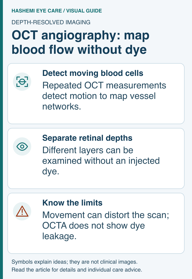
Visual guide: OCT angiography: map blood flow without dye
Read this visual as text
Detect moving blood cells: Repeated OCT measurements detect motion to map vessel networks. Separate retinal depths: Different layers can be examined without an injected dye. Know the limits: Movement can distort the scan; OCTA does not show dye leakage.
How It Works
- Uses OCT technology
- Detects motion of blood cells
- Creates maps of blood vessel networks
- Different depths can be analyzed separately
Why It's Done
- Evaluate optic nerve blood flow
- Detect vascular abnormalities
- Assess macular conditions
- Monitor glaucoma
- Research applications in neuro-ophthalmology
Advantages Over Fluorescein Angiography
| OCTA | Fluorescein Angiography |
|---|---|
| No dye injection | Requires IV dye |
| Can be repeated easily | Side effects from dye |
| Depth-resolved images | Surface view mainly |
| Quick scan | Longer procedure |
| No injected-dye reaction risk | Possible reaction to injected dye |
What to Expect
- Same setup as regular OCT
- Chin on rest, focus on target
- Multiple scans acquired
- Computer processes images
- Takes 5-10 minutes
Limitations
- Limited field of view
- Motion artifacts can affect quality
- Cannot show leakage (unlike FA)
- Technology still evolving
- Interpretation requires expertise
In Neuro-Ophthalmology
- Research tool for optic nerve conditions
- Assess papilledema
- Evaluate ischemic conditions
- Growing applications
References
- American Academy of Ophthalmology. Beginners Guide to OCT-A.
Medical Disclaimer: This information is for educational purposes only and does not replace professional medical advice.