

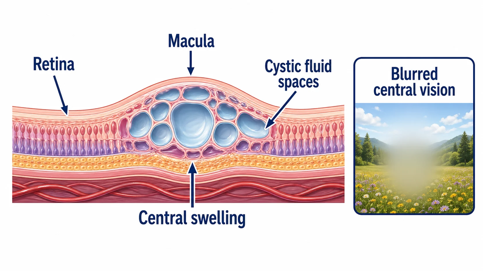
Macular edema is fluid swelling in the central retina. It can make straight lines look bent and cause blurred vision or central vision loss, affecting reading and faces. Diabetes, blocked retinal veins, inflammation, surgery, and wet AMD are common causes. OCT measures the swelling and helps guide treatment.
Key takeaways
- This is swelling of the central retina, not a separate disease by itself; something is making the macula leak or hold fluid
- Diabetes leads the list, but vein occlusions, eye surgery, inflammation, wet AMD, and medication effects also show up
- Vision may look blurry, wavy, faded, or hard to center on, especially for reading and faces
- OCT is the primary imaging test because it shows the swollen retinal layers in cross-section
- Treatment follows the cause: anti-VEGF injections, steroids, anti-inflammatory drops, laser, or tighter control of the underlying disease
- Old swelling can injure the macula, so timing matters; waiting for months is rarely a good plan
Questions about your diagnosis? Contact our office
Understanding Macular Edema
The macula provides sharp central vision and color detail. Normally, the blood-retinal barrier controls fluid moving into and out of the retina. When it leaks, fluid can distort the retinal layers and form small pockets called cystoid spaces, or cystoid macular edema (CME).
VEGF promotes vessel leakage; inflammatory signals also contribute, particularly after surgery or with uveitis. Longstanding swelling can permanently damage the light-sensing cells.
Inner vs. Outer Blood-Retinal Barrier
- Inner barrier: tight junctions in retinal blood vessels; breakdown occurs in diabetes and vein occlusions.
- Outer barrier: retinal pigment epithelium (RPE); disruption can occur in inflammatory disease and central serous chorioretinopathy.
Causes
Macular edema is not a disease in itself but rather a consequence of an underlying condition. Many different eye and systemic diseases can cause it.
Diabetic Macular Edema
Diabetic macular edema (DME) develops when damaged vessels leak into the macula. It can occur at any stage of diabetic retinopathy, in one or both eyes, and may initially cause no noticeable symptoms.
Approximately 7-10% of people with diabetes develop DME over their lifetime. Risk increases with longer disease duration and poor blood sugar control.
Retinal Vein Occlusion
Blockage of the retinal veins causes blood to back up, increasing pressure in the capillaries and leading to leakage and edema. Macular edema is the most common cause of vision loss in both:
- Central retinal vein occlusion (CRVO) - blockage of the main retinal vein, causing widespread edema and hemorrhage
- Branch retinal vein occlusion (BRVO) - blockage of a smaller branch vein, causing edema in the affected sector
Vein occlusion-related macular edema often develops suddenly and may be associated with risk factors such as hypertension, glaucoma, and cardiovascular disease.
Post-Surgical (Irvine-Gass Syndrome)
Inflammation after eye surgery, especially cataract surgery, can disrupt the blood-retinal barrier. This is called Irvine-Gass syndrome or pseudophakic CME and typically appears 4-12 weeks after surgery.
Clinically significant swelling occurs in approximately 1-2% of cataract operations; OCT can detect milder cases. Diabetes, uveitis, epiretinal membrane, and surgical complications increase risk.
Uveitis and Inflammatory Disease
Chronic or recurring uveitis can cause swelling that is difficult to control and may return with inflammatory flares. Associated conditions include sarcoidosis, Behcet disease, juvenile idiopathic arthritis, multiple sclerosis-associated intermediate uveitis, and Vogt-Koyanagi-Harada disease.
Retinal Vascular Disease
Other retinal vascular diseases can cause macular edema through similar mechanisms of vascular leakage. These include radiation retinopathy, retinal telangiectasia (such as Coats disease and macular telangiectasia type 2), and retinal artery macroaneurysm. In each case, abnormal blood vessels leak fluid into the macular region.
Other Causes
- Medication-related: Topical prostaglandin analogs used for glaucoma, certain chemotherapy agents, and fingolimod (used for multiple sclerosis) can cause or worsen macular edema.
- Epiretinal membrane: Tractional forces from a membrane on the retinal surface can contribute to edema.
- Age-related macular degeneration: Wet AMD involves fluid and blood leaking from abnormal new blood vessels under the retina.
Symptoms
Macular edema may develop gradually or appear suddenly, depending on the underlying cause. Symptoms primarily affect central vision.
Central Vision Changes
- Blurred central vision - the most common and often earliest symptom, ranging from mild haziness to significant loss of detail
- Difficulty reading - small print may become hard to see even with glasses
- Trouble recognizing faces - facial features may appear unclear
Visual Distortion
- Metamorphopsia - straight lines appear wavy, bent, or irregular; this occurs because the swollen macula physically distorts the photoreceptor arrangement
- Micropsia - objects may appear smaller than they actually are
- Macropsia - less commonly, objects may appear larger
Color and Contrast
- Decreased color perception - colors may appear washed out, faded, or slightly different between the two eyes
- Reduced contrast sensitivity - difficulty distinguishing objects from their background, especially in low light
Progression Pattern
In diabetic macular edema, symptoms often develop gradually over weeks to months. In vein occlusions, onset is typically sudden. Post-surgical macular edema usually appears weeks after the procedure. In many cases, the unaffected eye compensates, and patients may not notice mild edema until it is detected on examination.
Monitor your vision regularly. Because macular edema can develop gradually and one eye may compensate for the other, check each eye separately by covering the opposite eye. If you notice any new blurring, distortion, or difficulty reading, schedule an eye examination promptly. Early detection leads to better treatment outcomes.
Diagnosis
Accurate diagnosis of macular edema requires a combination of clinical examination and imaging. Identifying the underlying cause is equally important for guiding treatment.
Visual Acuity Testing
A visual acuity test measures your central vision and establishes a baseline. Best-corrected visual acuity (BCVA) is recorded and used to track treatment response. Vision may range from normal (if edema is mild or outside the foveal center) to severely reduced.
Optical Coherence Tomography (OCT)
OCT is a quick, painless scan showing the retina in cross-section. It measures thickness, locates fluid pockets or fluid beneath the retina, and checks the light-sensing layers for damage.
Comparing scans over time helps your ophthalmologist judge treatment response. OCT is often repeated at follow-up visits, alongside vision testing.
Fluorescein Angiography
Fluorescein angiography photographs retinal vessels after dye is injected into an arm vein. It shows where vessels leak, whether leakage is focal or widespread, and where blood flow is poor. These findings can guide laser treatment.
Fundoscopic Examination
A dilated eye examination checks for retinal thickening, fluid-related changes, yellow lipid deposits called hard exudates, and signs of the underlying disease, such as microaneurysms, bleeding, or cotton-wool spots from poor blood supply.
Treatment
Treatment of macular edema depends on the underlying cause, severity of edema, visual acuity, and individual patient factors. The goals are to reduce swelling, prevent further damage, and improve or stabilize vision.
Anti-VEGF Injections
Anti-VEGF injections reduce vessel leakage and are commonly used for diabetic macular edema, retinal vein occlusion, and wet AMD. Post-surgical or inflammatory swelling may need a different approach.
Options include ranibizumab, aflibercept, faricimab, and off-label bevacizumab. The choice and interval depend on the diagnosis, formulation, OCT findings, vision, and response; there is no single schedule for every type of edema.
The eye is numbed and cleaned before this brief office procedure. Treatment often starts with monthly visits, with intervals adjusted afterward. Some patients need treatment for years.
In the DRCR.net Protocol T trial, aflibercept, bevacizumab, and ranibizumab all improved DME-related vision loss. At one year, aflibercept gave greater benefit in eyes starting at 20/50 or worse.
After an injection, contact your retina team immediately for increasing pain, redness, light sensitivity, new floaters, or reduced vision. These symptoms need assessment rather than waiting for the next routine visit.
Corticosteroid Therapy
Corticosteroids can help inflammatory edema and selected cases that respond poorly to anti-VEGF treatment.
- Dexamethasone implant (Ozurdex): releases medication over about 3-6 months; used for DME, vein-occlusion edema, and non-infectious posterior uveitis.
- Fluocinolone implant (Iluvien): releases medication for up to 36 months; approved for DME after prior steroid treatment without a significant pressure rise.
Steroids can raise eye pressure and accelerate cataracts, so follow-up must include pressure checks. An injection also carries a rare risk of serious infection inside the eye (endophthalmitis).
Laser Photocoagulation
Laser is sometimes added for persistent leakage or when injections are unsuitable or unavailable.
- Focal laser targets individual leaking microaneurysms.
- Grid laser treats areas of more widespread thickening.
- The modified ETDRS technique is used when laser is chosen for diabetic edema.
Laser may reduce the need for injections in selected cases; it does not replace them for every patient.
Treating the Underlying Cause
Addressing the root cause of macular edema is essential for long-term success:
- Diabetes: Work with your diabetes clinician on an individualized A1c target, blood pressure and cholesterol management to reduce diabetic eye risks
- Retinal vein occlusion: Managing cardiovascular risk factors (hypertension, hyperlipidemia) and evaluating for underlying conditions
- Uveitis: Controlling inflammation with systemic immunomodulatory therapy, topical and periocular steroids
- Medication-related: The prescriber may discontinue or substitute a suspected medicine when appropriate; do not stop it independently
NSAIDs for Post-Surgical Macular Edema
After surgery, NSAID eye drops may be used alone or with prescribed steroid drops. Examples include ketorolac (Acular), nepafenac (Nevanac, Ilevro), and bromfenac (Prolensa).
The surgeon sets the start date and duration; courses often continue for 4-6 weeks after surgery. Persistent swelling may need injections or another treatment. Do not change or stop drops without discussing the plan with your eye team.
Prognosis
The prognosis for macular edema varies widely depending on the underlying cause, duration of edema before treatment, and response to therapy.
Favorable Prognostic Factors
- Early detection and treatment before chronic structural damage occurs
- Good baseline visual acuity at the start of treatment
- Prompt response to anti-VEGF therapy (reduction in edema after first few injections)
- Effective management of the underlying condition (well-controlled diabetes, resolved inflammation)
- Absence of macular ischemia on fluorescein angiography
Less Favorable Prognostic Factors
- Chronic, longstanding edema with photoreceptor damage
- Significant macular ischemia (poor blood flow to the macula)
- Hard exudate deposits in the foveal center
- Disruption of the outer retinal layers (ellipsoid zone) on OCT
- Poor control of underlying systemic disease
General Outcomes
With modern anti-VEGF therapy, many patients experience significant visual improvement. In clinical trials for diabetic macular edema, approximately 30-40% of treated patients gained 15 or more letters (3 lines) of vision on an eye chart after one year of treatment. Most patients experience at least stabilization of vision with appropriate treatment.
Post-surgical macular edema has an excellent prognosis; most cases resolve with topical NSAID therapy, and vision typically returns to expected post-operative levels. Uveitic macular edema can be more challenging, with recurrence common when inflammation flares.
Prevention
While macular edema cannot always be prevented, several strategies significantly reduce the risk:
For People with Diabetes
- Maintain tight blood sugar control - the Diabetes Control and Complications Trial (DCCT) and UK Prospective Diabetes Study (UKPDS) showed that intensive glycemic control reduces the risk of diabetic eye complications by up to 76%
- Control blood pressure - hypertension worsens diabetic retinopathy and macular edema
- Manage cholesterol - elevated lipids contribute to hard exudate deposits in the macula
- Get annual dilated eye exams - early detection allows treatment before significant vision loss
- Do not skip appointments - consistent monitoring is key to catching changes early
For Everyone
- Report new vision changes promptly - blurred or distorted central vision deserves evaluation
- Follow post-operative instructions carefully - use prescribed anti-inflammatory drops after eye surgery as directed
- Manage cardiovascular risk factors - high blood pressure, high cholesterol, and smoking increase the risk of retinal vascular disease
- Protect your eyes from injury - ocular trauma can trigger inflammation and edema
Frequently Asked Questions
Is macular edema the same as macular degeneration?
No. Edema means fluid swelling and has many causes. Macular degeneration is an age-related disease involving drusen or abnormal new vessels. Wet AMD can cause macular fluid, so the conditions can overlap.
How many injections will I need?
It depends on the cause and your response. Initial monthly treatment may be followed by longer intervals, but some eyes need injections for several years. OCT and vision checks guide each decision; do not skip a visit because vision seems stable.
Are anti-VEGF injections painful?
Numbing drops usually limit discomfort to brief pressure. Mild grittiness or a small surface red spot can occur afterward. Increasing pain, light sensitivity, or reduced vision needs immediate contact with your retina team.
Can macular edema come back after treatment?
Yes, especially when diabetes or inflammation remains active. Continued monitoring helps detect recurrence and adjust treatment. Discuss changes with your ophthalmologist rather than stopping treatment when vision improves.
Will my vision return to normal?
Many patients improve, especially with early treatment. Longstanding swelling or poor blood flow can permanently damage the macula, limiting recovery even after the fluid resolves.
References
- American Society of Retina Specialists. Intravitreal Injections.
- American Academy of Ophthalmology. Macular Edema.
- National Eye Institute. Macular Edema.
- Diabetic Retinopathy Clinical Research Network (DRCR.net). Protocol T: Comparative Effectiveness of Anti-VEGF Agents for Diabetic Macular Edema. N Engl J Med. 2015;372(13):1193-1203.
- Schmidt-Erfurth U, Garcia-Arumi J, Bandello F, et al. Guidelines for the Management of Diabetic Macular Edema by the European Society of Retina Specialists (EURETINA). Ophthalmologica. 2017;237(4):185-222.
- American Diabetes Association. Glycemic goals: Standards of Care in Diabetes—2026.
- Yonekawa Y, Kim IK. Pseudophakic Cystoid Macular Edema. Curr Opin Ophthalmol. 2012;23(1):26-32.
- Daruich A, Matet A, Moulin A, et al. Mechanisms of Macular Edema: Beyond the Surface. Prog Retin Eye Res. 2018;63:20-68.
Medical Disclaimer: This information is for educational purposes only and does not replace professional medical advice, diagnosis, or treatment. If you are experiencing blurred or distorted central vision, schedule an appointment with your ophthalmologist promptly.