

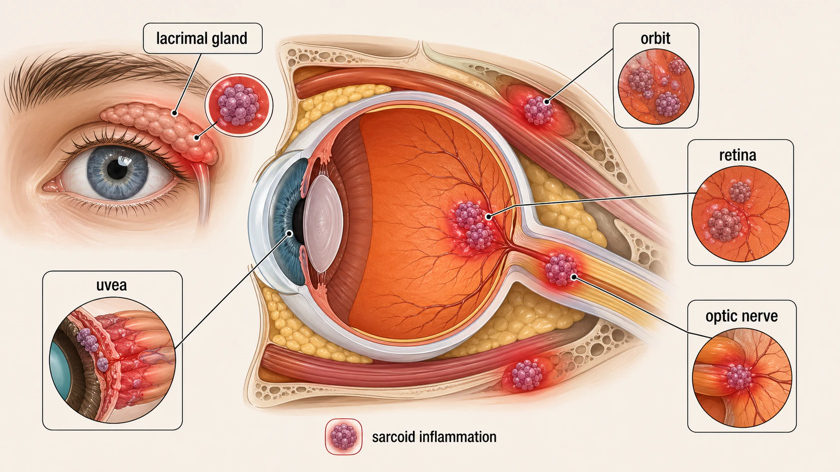
Ocular sarcoidosis may be the first manifestation of systemic sarcoidosis. A red, light-sensitive eye, new floaters, dry eye from lacrimal gland involvement, or optic nerve inflammation can occur when sarcoid granulomas and related inflammation involve ocular tissues. Ocular sarcoidosis can affect nearly any eye structure, though uveitis is the most common manifestation. Roughly 25-50% of people with sarcoidosis have eye involvement. Early recognition and control of inflammation help reduce the risk of preventable vision loss.
Key takeaways
- Eye symptoms may be the first manifestation of sarcoidosis, even before lung or skin findings are obvious
- Anterior uveitis is the usual pattern, with eye pain, redness, and photophobia
- The workup ties the eye to the rest of the body through eye exam findings, blood tests, chest imaging, and IWOS criteria
- Steroids often calm flares, but chronic disease may need methotrexate, azathioprine, mycophenolate, or another steroid-sparing medication
- Complications are not rare: cataracts, glaucoma, and cystoid macular edema need monitoring
- The course can relapse, so follow-up is an important part of treatment
Questions about your diagnosis? Contact our office
Understanding Sarcoidosis
Sarcoidosis causes clusters of inflammatory cells called non-caseating granulomas. It most often affects the lungs and lymph nodes but can involve the skin, heart, nervous system, eyes, and other organs.
The cause is unknown. An immune response to an environmental trigger in genetically susceptible people is one proposed explanation. Eye inflammation may occur before other signs of systemic disease are recognized.
How Sarcoidosis Affects the Eyes
Sarcoidosis can involve nearly every structure of the eye. The specific manifestations depend on which tissues are affected.
Anterior Uveitis
Anterior uveitis, affecting the iris and ciliary body, is the most common pattern. Findings on slit-lamp examination include:
- Mutton-fat keratic precipitates: large inflammatory deposits on the back of the cornea.
- Busacca and Koeppe nodules: small inflammatory nodules on the iris or pupil margin.
- Cells and flare: inflammatory cells and protein in the front chamber.
- Posterior synechiae: iris-lens adhesions that can affect the pupil.
Inflammation may be acute or persistent. Chronic disease is more likely to cause complications.
Intermediate Uveitis
Inflammation in the vitreous and nearby peripheral retina may produce snowballs (floating inflammatory clumps), snowbanking (deposits near the pars plana), and vitreous haze causing blurred vision or floaters. It may coexist with anterior or posterior inflammation.
Posterior Uveitis
Inflammation in the retina, choroid, or retinal vessels may cause:
- Periphlebitis ("candle-wax drippings"): inflammation and deposits along retinal veins.
- Choroidal granulomas or multifocal choroiditis: nodules or inflammatory lesions in the layer beneath the retina.
- Retinal vasculitis: vessel inflammation that can impair blood flow.
- Cystoid macular edema: central retinal swelling, an important cause of vision loss.
- Inflammatory cells and haze in the vitreous.
Optic Nerve Involvement
Sarcoidosis can cause disc edema, optic nerve infiltration, or a granuloma around the disc, with potentially serious vision loss. It can resemble optic neuritis or papilledema and may occur as part of neurosarcoidosis.
Lacrimal Gland Involvement
The tear glands may enlarge painlessly, often on both sides. Gland damage can reduce tear production and cause dry eye. This may be the first sign of sarcoidosis.
Orbital Involvement
Less often, inflammation or a granulomatous mass in the eye socket causes proptosis or restricted eye movements with double vision. It can resemble orbital inflammatory disease or lymphoma. Eyelid nodules may also occur.
Symptoms
The symptoms of ocular sarcoidosis depend on which parts of the eye are affected. Some patients have prominent symptoms, while others have minimal complaints despite active inflammation.
Common Symptoms
- Eye pain: aching or deep pain, particularly with anterior uveitis
- Photophobia: sensitivity to light, often severe with anterior uveitis
- Redness: a red or pink eye, especially circumlimbal (around the cornea)
- Blurred vision: from inflammation, macular edema, or vitreous haze
- Floaters: from vitreous cells or inflammatory debris
- Dry eyes: from lacrimal gland involvement
- Double vision: from orbital or cranial nerve involvement
Less Common Symptoms
- Decreased color vision: if the optic nerve is involved
- Visual field loss: from optic neuropathy or glaucoma
- Painless vision loss: may occur with posterior uveitis or chronic macular edema
- Tearing-paradoxically, early lacrimal gland inflammation can cause excess tearing before dry eye develops
Ocular sarcoidosis can be present with minimal or no symptoms. Some patients with active posterior inflammation or early glaucoma have no complaints until significant damage has occurred. This is why regular ophthalmological screening is often recommended for patients diagnosed with systemic sarcoidosis.
Diagnosis
Diagnosing ocular sarcoidosis requires a combination of clinical eye examination, laboratory testing, and imaging. The diagnosis can be challenging because sarcoidosis can mimic many other inflammatory conditions.
Slit-Lamp Examination
A slit-lamp exam checks for corneal deposits, inflammatory cells, iris nodules, adhesions, and vitreous haze. The clinician also checks the lens for cataracts and measures pressure because inflammation or treatment can contribute to glaucoma.
Optical Coherence Tomography (OCT)
OCT measures retinal thickness and detects macular fluid, surface membranes, and other structural changes. Repeat scans help assess treatment response and optic nerve swelling.
Fluorescein Angiography
Fluorescein angiography photographs retinal vessels after an arm-vein dye injection. It can show inflamed or leaking veins, macular swelling, optic disc leakage, and areas of poor blood flow.
Blood Tests
Blood tests support the diagnosis and check organ involvement; none confirms sarcoidosis alone.
- ACE and lysozyme may rise with granuloma activity. Normal ACE does not rule out sarcoidosis.
- Blood count may show low lymphocytes.
- Liver, kidney, and calcium tests check systemic effects; calcium can be elevated.
- ESR and CRP measure inflammation.
Chest Imaging
A chest X-ray may show enlarged lymph nodes at the lung roots (bilateral hilar lymphadenopathy). A chest CT scan gives more detail when needed. Lung findings help connect eye inflammation with systemic sarcoidosis.
MRI
MRI of the brain and orbits is indicated when there is:
- Suspected optic nerve involvement
- Orbital disease causing proptosis or restricted eye movement
- Concern for neurosarcoidosis (cranial nerve palsies, meningitis, or brain lesions)
- Unexplained disc edema to differentiate from other causes
Biopsy
A biopsy showing non-caseating granulomas provides strong diagnostic support. The team may sample an accessible lymph node, skin or conjunctival nodule, enlarged lacrimal gland, or lung tissue.
Biopsy is particularly useful when the diagnosis remains uncertain; the choice depends on which site can be sampled safely.
International Workshop on Ocular Sarcoidosis (IWOS) Diagnostic Criteria
The revised 2019 IWOS framework combines eye findings, systemic investigations, and biopsy results after other causes of granulomatous uveitis are excluded. Specialists use the complete criteria to judge how strongly the evidence supports sarcoidosis; an abnormal blood test alone is not a diagnosis.
Treatment
Treatment of ocular sarcoidosis aims to control inflammation, prevent complications, and preserve vision. The approach is tailored to the severity and location of eye involvement.
Corticosteroids
Corticosteroids are the first-line treatment for ocular sarcoidosis and are used in various forms depending on the location and severity of inflammation.
Topical corticosteroids (eye drops):
- First-line for anterior uveitis
- Prednisolone acetate 1% or difluprednate 0.05% are commonly used
- Your ophthalmologist sets the frequency and taper based on the examination
- Follow-up checks eye pressure and monitors for steroid-induced glaucoma and cataracts
- Do not restart or self-refill steroid drops. They can worsen herpes simplex and other eye infections; a new flare needs an examination
Periocular corticosteroid injections:
- Sub-Tenon or orbital floor injections of triamcinolone acetonide
- Used for intermediate or posterior uveitis unresponsive to topical drops
- Provide sustained local anti-inflammatory effect
- Risk of elevated intraocular pressure
Intravitreal corticosteroid injections:
- Triamcinolone acetonide or dexamethasone implant (Ozurdex)
- Targeted delivery to the posterior segment for macular edema or posterior uveitis
- Dexamethasone implant provides sustained release over approximately 3-6 months
- Risks include elevated intraocular pressure and cataract progression
Systemic corticosteroids:
- Oral prednisone is used for severe, bilateral, or sight-threatening ocular sarcoidosis
- Typical starting dose is 0.5-1 mg/kg/day, tapered gradually over weeks to months
- Also treats concurrent systemic sarcoidosis
- IV steroids (intravenous methylprednisolone) may be used for severe optic neuropathy or acute sight-threatening inflammation
- Long-term systemic steroids carry significant side effects (weight gain, diabetes, osteoporosis, adrenal suppression)
Steroid-Sparing Immunosuppressive Agents
For recurring or persistent inflammation, steroid-sparing medicines can reduce the need for long-term prednisone:
- Methotrexate: the most commonly used steroid-sparing agent for ocular sarcoidosis. Given weekly (oral or injection), it is effective in controlling uveitis and reducing steroid dependence. Requires monitoring of liver function and blood counts
- Azathioprine: an alternative immunosuppressant used when methotrexate is not tolerated. Requires TPMT enzyme testing before initiation and regular blood monitoring
- Mycophenolate mofetil (CellCept): another effective steroid-sparing option, often used for posterior uveitis and macular edema. Generally well-tolerated but requires blood count monitoring
These agents typically take 2-3 months to reach full effectiveness, during which time corticosteroids bridge the gap.
Biologic Agents
Biologics may be considered when steroids and conventional medicines do not control inflammation:
- Adalimumab (Humira): an anti-TNF-alpha monoclonal antibody approved for non-infectious intermediate, posterior, and panuveitis. Given as subcutaneous injections every two weeks. Has strong evidence for uveitis management
- Infliximab (Remicade): another anti-TNF-alpha agent, given as intravenous infusions. Often effective for severe or refractory ocular and neurosarcoidosis
- Rituximab: an anti-CD20 monoclonal antibody that depletes B cells. Used in select refractory cases, particularly when other biologics have failed
Biologic agents require screening for tuberculosis and hepatitis before initiation and carry risks of serious infection and other immune-related complications.
Treatment is often long-term. Ocular sarcoidosis frequently follows a chronic, relapsing course. Many patients require years of immunosuppressive therapy. The goal is to achieve and maintain remission using the lowest effective medication dose, minimizing side effects while preventing sight-threatening inflammation.
Complications
Without adequate treatment, ocular sarcoidosis can lead to several sight-threatening complications. Even with treatment, some complications may develop, requiring additional management.
Cataracts
Cataracts are a common complication, resulting from both chronic intraocular inflammation and prolonged corticosteroid use. Cataract surgery can be performed once inflammation is controlled for at least 3 months, though the surgical management of cataract in uveitis patients requires special considerations.
Glaucoma
Glaucoma may result from steroid-related pressure elevation, inflammatory debris blocking drainage, or iris-lens adhesions that close the drainage angle. Pressure checks are needed even when the eye feels comfortable.
Cystoid Macular Edema (CME)
Fluid accumulation in the macula is one of the most common causes of vision loss in ocular sarcoidosis. CME can persist despite control of visible inflammation and may require targeted intravitreal treatment.
Band Keratopathy
Calcium deposits in the superficial cornea (band keratopathy) can develop in chronic anterior uveitis, causing discomfort and reduced vision. Treatment involves chelation with EDTA solution.
Optic Atrophy
Untreated or severe optic nerve involvement can lead to optic atrophy, irreversible damage to the optic nerve with permanent vision loss. This underscores the importance of aggressive treatment when the optic nerve is involved.
Other Complications
- Epiretinal membrane formation: scar tissue on the retinal surface causing distortion
- Retinal neovascularization: abnormal new blood vessel growth from chronic ischemia
- Vitreous hemorrhage: bleeding from neovascularization
- Hypotony: low eye pressure from ciliary body damage due to chronic inflammation
Prognosis
The prognosis for ocular sarcoidosis is generally favorable with appropriate treatment, but outcomes vary depending on the severity and chronicity of the disease.
Favorable Factors
Earlier diagnosis, prompt treatment, an acute anterior-uveitis pattern, and consistent medication use and follow-up are associated with better outcomes. Absence of optic nerve involvement is also favorable.
Unfavorable Factors
Persistent posterior inflammation, recurrent macular edema, delayed treatment of optic nerve disease, glaucoma, and difficulty tolerating steroid-sparing medication can limit recovery.
Long-Term Outlook
- Many patients with ocular sarcoidosis achieve good visual outcomes with treatment
- Approximately two-thirds of patients with sarcoid uveitis maintain vision of 20/40 or better
- However, up to 10-15% of patients may develop significant visual impairment despite treatment
- The disease often follows a relapsing-remitting course, with flares requiring treatment adjustments
- Some patients achieve sustained remission and can eventually discontinue therapy, though this may take years
- Multidisciplinary management with pulmonology or rheumatology is often needed for systemic disease management
Frequently Asked Questions
Can ocular sarcoidosis cause permanent vision loss?
Yes. Glaucoma, macular edema, optic nerve damage, and cataracts can reduce vision. Treatment and regular checks help protect useful vision, including when symptoms seem mild.
Will I need to take medications for life?
Not everyone does. A single episode may settle, while recurring disease can need treatment for years. Your eye and systemic-care teams aim for the lowest effective dose and consider tapering when inflammation is controlled.
I have sarcoidosis but no eye symptoms. Should I see an ophthalmologist?
Yes. Arrange a baseline slit-lamp examination and dilated eye examination, because inflammation or glaucoma may be silent. Follow-up is often every 6-12 months, sooner if eye involvement is found; your clinician sets the interval.
Is ocular sarcoidosis contagious?
No. Sarcoidosis is an immune-mediated condition and does not spread from person to person. Its trigger remains unknown.
What is the difference between sarcoid uveitis and other types of uveitis?
Sarcoid uveitis often has a granulomatous pattern: large corneal deposits, iris nodules, or characteristic retinal-vein inflammation. These findings guide the workup, but tuberculosis and syphilis can produce similar inflammation and must be considered.
References
- DailyMed. PRED FORTE (prednisolone acetate ophthalmic suspension) prescribing information.
- American Academy of Ophthalmology. Uveitis.
- Mochizuki M, Smith JR, Takase H, et al. Revised criteria of International Workshop on Ocular Sarcoidosis (IWOS) for the diagnosis of ocular sarcoidosis. Br J Ophthalmol. 2019;103(10):1418-1422.
- American Thoracic Society. Statement on sarcoidosis. Am J Respir Crit Care Med. 1999;160(2):736-755.
- Pasadhika S, Rosenbaum JT. Ocular sarcoidosis. Clin Chest Med. 2015;36(4):669-683.
- Rothova A. Ocular involvement in sarcoidosis. Br J Ophthalmol. 2000;84(1):110-116.
- National Eye Institute. Uveitis.
Medical Disclaimer: This information is for educational purposes only and does not replace professional medical advice, diagnosis, or treatment. If you have symptoms of ocular sarcoidosis or have been diagnosed with sarcoidosis, consult with an ophthalmologist for comprehensive eye evaluation.