

This case report describes a 48-year-old man who developed acute vision loss in the right eye following head trauma from a car accident, found to have a ruptured retinal arterial macroaneurysm (RAM) with associated retinal hemorrhage and exudate. Published in the Journal of Ophthalmology Research Reviews & Reports in 2021, the case illustrates how trauma can precipitate rupture of a pre-existing retinal macroaneurysm and emphasizes the importance of thorough retinal evaluation in patients with post-traumatic visual symptoms.
Key takeaways
Key Findings
- Retinal arterial macroaneurysm (RAM) ruptured following head trauma from a car accident in a 48-year-old man with no prior medical history
- Vision was reduced to 20/100 in the right eye with impaired color vision (6/11 OD), while the left eye remained 20/20
- Dilated exam revealed inferotemporal retinal hemorrhage with exudate in the right eye, consistent with ruptured RAM
- Visual field testing showed a superior nasal defect in the right eye, correlating anatomically with the inferotemporal hemorrhage location
- OCT of the retina and optic nerve were within normal limits, and no RAPD was present
- Management and recovery depend on the hemorrhage location and effect on the macula; this report does not document a final visual outcome
Questions about your diagnosis? Contact our office
Visual guide: Retinal bleeding after head trauma
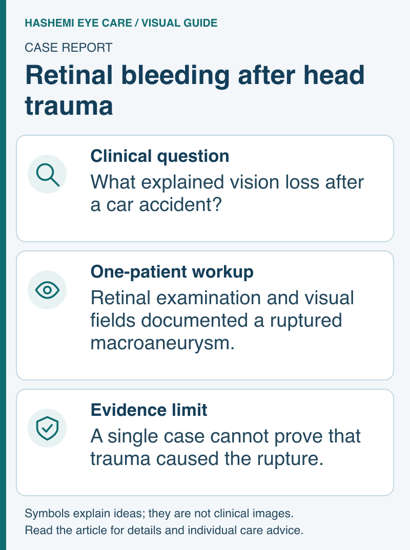
Read this visual as text
Background
Retinal arterial macroaneurysm (RAM) is an acquired, focal dilation of a retinal artery that can present with retinal hemorrhage or exudation. It is most commonly associated with systemic hypertension, arteriosclerosis, and cardiovascular disease. RAM can present with a wide variety of clinical appearances, leading to frequent misdiagnosis as other conditions including Coats disease, von Hippel-Lindau disease, retinal vein occlusion, and diabetic retinopathy.
Fluorescein angiography is one of the most useful diagnostic tests for confirming RAM. Management depends on the clinical findings and associated complications. Some RAMs thrombose or scar without treatment, but visual recovery depends on retinal damage and the location of bleeding or fluid. However, complications such as macular hole formation have been reported secondary to ruptured RAM.
While RAM is typically seen in patients with underlying vascular risk factors, trauma can serve as a precipitating event for rupture of an existing macroaneurysm. This case is notable because the patient was otherwise healthy with no known medical history, and the rupture occurred in the setting of head trauma from a motor vehicle accident.
The Patient
A 48-year-old Hispanic man presented to the neuro-ophthalmology clinic with:
- Blurry vision in the right eye
- History of head trauma from a car accident in March 2020
- No loss of consciousness at the time of the accident
- Vision loss in the right eye noticed the day after the car accident
- No associated headaches, diplopia, or other eye-related symptoms
- No past medical history (no hypertension, diabetes, or cardiovascular disease)
Diagnostic Workup
Clinical Examination
- Visual acuity: 20/100 OD (right eye), 20/20 OS (left eye)
- Intraocular pressure: 18 mmHg in both eyes
- Color vision test: 6/11 OD (reduced), 11/11 OS (normal)
- Ocular motility: No nystagmus or intrusion in saccadic or pursuit movements
- Pupil exam: Normal pupillary reaction to light; no relative afferent pupillary defect (RAPD)
Retinal Findings
- Dilated fundus examination: Inferotemporal retinal hemorrhage associated with exudate in the right eye; normal retinal exam in the left eye
- OCT of the retina and optic nerve: Within normal limits
- Fundus photography: Confirmed inferotemporal hemorrhage with exudate in the right eye
Visual Field Testing
- Visual field test: Superior nasal defect in the right eye, anatomically consistent with the inferotemporal retinal hemorrhage location
Results
The clinical presentation -- unilateral retinal hemorrhage with exudate following head trauma in an otherwise healthy patient -- was consistent with a ruptured retinal arterial macroaneurysm. Key diagnostic correlations included:
- Inferotemporal hemorrhage location correlated precisely with the superior nasal visual field defect, following the expected retinotopic mapping
- Absence of RAPD indicated that the optic nerve was not significantly affected
- Normal OCT suggested the hemorrhage and exudate did not cause significant structural macular changes at the time of imaging
- Unilateral involvement (right eye only) is typical for RAM
The temporal relationship between the car accident and onset of visual symptoms led the authors to suggest that trauma precipitated rupture; a single case cannot prove that causal relationship.
Clinical Significance
This case highlights several important clinical points:
- Trauma can trigger rupture of a pre-existing retinal arterial macroaneurysm, even in patients without known vascular risk factors
- Post-traumatic visual complaints require thorough retinal evaluation, including dilated fundus examination and visual field testing, to rule out retinal pathology
- RAM is often misdiagnosed due to its variable clinical presentation; differential diagnosis includes Coats disease, von Hippel-Lindau disease, retinal vein occlusion, and diabetic retinopathy
- Prognosis is individualized -- spontaneous closure can occur, but macular damage or persistent bleeding can limit visual recovery
- Patients with RAM should be monitored for systemic conditions including hypertension, hyperlipidemia, and arteriosclerosis, even when initially presenting without these risk factors
Management Guidelines
A retina specialist decides whether observation or intervention is appropriate and sets the follow-up interval from vision, bleeding, fluid and macular involvement. New post-traumatic vision loss needs prompt assessment rather than waiting for an observation schedule from a case report. Macular hole formation is one reported complication.
Clinical Pearl: When evaluating a patient with new vision loss after head trauma, do not assume that all visual symptoms are related to traumatic brain injury or post-concussive syndrome. A thorough dilated retinal examination is essential to rule out retinal vascular pathology such as ruptured macroaneurysm, which can be precipitated by trauma and may require different monitoring and follow-up.
Systemic Screening: Even when retinal arterial macroaneurysm is found in a patient without known vascular risk factors, screening for hypertension, hyperlipidemia, and arteriosclerosis is recommended. RAM is typically associated with systemic vascular disease, and the retinal finding may be the first manifestation of an undiagnosed systemic condition.
Citation
Naser M, Hashemi N. Macroaneurysm Rupture after Car Accident. Journal of Ophthalmology Research Reviews & Reports. 2021;2(1):1-2. SRC/JORRR/109.
References
Original Publication
- Naser M, Hashemi N. Macroaneurysm Rupture after Car Accident. Journal of Ophthalmology Research Reviews & Reports. 2021;2(1):1-2. SRC/JORRR/109.
Key References Cited in the Original Study
- Kester E, Walker E. Retinal arterial macroaneurysm causing multilevel retinal hemorrhage. Optometry. 2009;80:425-430.
- Rabb M, Gagliano D, Teske M. Retinal arterial macroaneurysm. Surv Ophthalmol. 1988;33:73-96.
- Gurwood AS, Nicholson CR. Retinal arterial macroaneurysm: a case report. J Am Optom Assoc. 1998;69:41-48.
- Sato R, Yasukawa T, Hirano Y, Ogura Y. Early-onset macular holes following ruptured retinal arterial macroaneurysms. Graefes Arch Clin Exp Ophthalmol. 2008;246:1779-1782.
Disclaimer: This page summarizes a peer-reviewed publication for educational purposes. It does not constitute medical advice. For clinical decisions, consult the original publication and a qualified healthcare provider.
Additional Treatment Context:
- Lam SS, et al. Clinical outcomes of RAM with vitreous hemorrhage managed by observation, anti-VEGF or vitrectomy. J Vitreoretin Dis. 2023.